Flap Anatomies
Flap Anatomies exist within the genre of 3D printed texts, inviting viewers and readers to participate in the movement of internal organs and various body parts. A flap anatomy can be defined as any paper-based printed image containing more than one layer, illustrating any part of human anatomy, often used for virtual autopsies and exploring human dissection. Other commonly used terms are “moveable books,” “fugitive sheets,” “pop-up books,” and “cut-out overlays.” While “flap anatomies” describe texts about biological concepts and functions, usage shifted over time, leading to a name shift as well. “Pop-up books” and “moveable books,” were terms used in later centuries, referring to more leisurely, fictional reading, targeted towards children. Starting with their first emergence in the early 16th century, flap anatomies have completely revolutionized the way the general public perceives the human body. Additionally, it generated a new and complex technology that allowed for advancement and exploration of the medical field, proving to be extremely beneficial for students, doctors, and teachers alike.
Interest in human anatomy grew immensely during the sixteenth century in Europe. This was during the bustling Renaissance period, in which major advancements were made in the fields of science, technology, and art. These scientific advancements also lead to scientific curiosities. As scientists chipped away at this biological mystique, they found that illustration was an efficient way to spread their knowledge to a broader audience. However, there was a common struggle and barrier in understanding: how to accurately depict the body’s three-dimensional structure on a flat piece of paper.[1] Although 2D images may be helpful, they also hindered complete comprehension. This problem was easily solved by flap anatomies, where superimposed sheets created multiple dimensions for viewers to analyze and understand.[2] By adding a sense of touch in addition to a sense of sight, the images became more realistic, and therefore easier to follow. Further, creating both visual and tactile aspects, these books are useful for both those who learn simply from sight, and those who benefit from more physical learning as well. Ironically, it was not only doctors and professionals that took an interest in the anatomical pamphlets. These flap illustrations actually attracted a much wider audience, seeking general education stemming from lack of knowledge or curiosity. [3] Simply, the public was unaware of how their bodies truly functioned, and what started as a technology for a niche group, actually transformed into a technology for a more general audience. The creation of flap anatomies answered many questions, solved many mysteries, and transformed Europe’s biological understanding.
The first ever anatomical fugitive sheet was Heinrich Vogtherr’s Strasbourg Edition, which was printed in Germany in 1538.[4] All of his sheets contained a “base sheet” with the printed outline of the body. Internal organs were printed on separate sheets, cut out, and then adhered to the base sheet at the top of each organ. Each organ had its name written in Latin, while the most important organs were mentioned in text that provided additional anatomical information. Interestingly, the male and female figures were printed from the same blocks and therefore, made from the same drawings.[5] It can be inferred that this was to keep production and retails costs at a low. However, it also revealed the similarities in the internal organs between men and women. Given that Vogtherr’s booklet was the first of its kind, many other printers, engravers, and illustrators copied his sheets in various editions, which were later colored and printed in multiple languages according to region. Vogtherr initiated a surge of anatomical sheets, created from superimposed woodcuts, that were published repeatedly in Germany, and eventually spread to other European nations.
As flap anatomies became increasingly more popular in multiple countries, and as advancements in printing shaped the industry, the technologies used to create these anatomical books shifted as well.
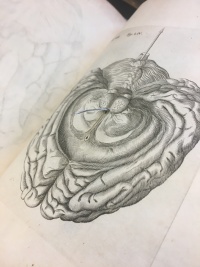
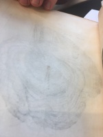
Flap anatomies produced during the 15th and 16th centuries had less intricate moving parts. Printers would often send instructions to binders regarding the cutting, gluing, and sewing of the flaps onto the base sheets.[6] This collaboration also contributed to the rapid spread of these technologies to other European countries. An example of the original, simple methodology of flap books is seen in René Descartes’, De Homine, an early flap anatomy created in 1664 in the Netherlands. Analyzing this book in the Rare Book & Manuscript Library at the University of Pennsylvania, there is at maximum of one delicate flap per image. The images display the front and back of one of the pages in the book. In this case, a small slit was cut into the base sheet, and the tiny flap, demonstrating a part of the brain, was put inside the slit, and adhered to the back of the page using glue or tape. As seen in the images, the flap now was able to stand on its own, and be moved in a somewhat free fashion. Viewers are better able to explore the functionality of this anatomy, and obtain a more realistic view of this part of the brain. However, this model is highly delicate, as the pieces of paper are thin and can be easily ripped or damaged. Additionally, since only glue and tape held the flap in the slit, often they would fall out and have to be replaced. Given such a delicate nature and extensive production process, flap anatomies at this time were not super common, and readership was limited to those whom were studying biology or in the medical field. These limitations initiated further invention and innovation in the production of flap anatomies.
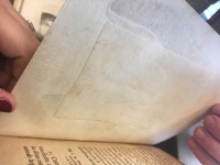
As technologies advanced, the methodologies for creating flap books advanced as well. In the early 1800s, printers and illustrators started to adhere the flaps all at the same place, which was usually the top of the image. One slit would be created and each individual flap would be inserted into the opening, making movement of the flaps sturdier.[7] Additionally, rather than leaving the flap simply glued down to the back of the page, printers started placing another sheet of paper over the glued or sewed flap, which is demonstrated by the image on the right. This also increased the durability of the flap book, because it decreased the chances of the flap falling out of the slit, since there was now paper to cover it. Both of these technologies were seen in George Spratt’s Obstetric Tables, which was produced in 1847.[8]
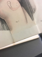
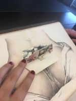
Spratt’s book also revealed another trend seen amongst many flap anatomies: the interest in the secrecy of the female body. During the Renaissance era, gender norms were being challenged and explored – specifically how the body, and the things you put on your body, defined whether you are male or female. Consequently, the public became extremely interested in the differences between the male and female bodies. Since many of the illustrations of the body were copied and reused, it was clear that men and women had many commonalities when it came to internal organs. However, the differences were often the ideas that were emphasized. As seen in the images, flap anatomies often explored biological processes that belong exclusively to women, such as the menstrual cycle or pregnancy.[9] The picture on the right was used to explain different trimesters of pregnancy: what the stomach should look like and how the fetus is developing. The picture on the left was used to explain the different hormones and organs at play when a woman is on her period. This interest, along with the wide spread use of flap anatomies and advancing technology, lead to an increasing knowledge of our anatomical functions and biological processes among the general public. As flap anatomies became mass-produced, and color images were introduced, these gender differences were continually highlighted and stressed. Since most individuals in the medical profession were men, these variances often lead to ideas about female inferiority, which then influenced perceptions of the general population.
The 19th and 20th centuries were considered the “Golden Age of Flap Books” due to advancements in printing that allowed for anatomical parts to be displayed in vivid, dramatic colors. [10]. Again, a color image allowed for a general increase in biological understanding, since it was more difficult to learn about overlapping pathways and body functions while the illustrations were solely in black and white. These later flap books also had different methodologies of creation due to news types of machines and printers. Each page was actually two sheets adhered together with an area of space between them designed to create room for all of the extra paper. This specific method was even sturdier than previous methods, due to the size of the tabs adhered between the layers. Additionally, flaps would often rise upon opening to a specific page of the book, without the necessary lifting of the reader.
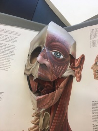
In these models, there also existed tabs that viewers could pull, that would cause for the movement of different flaps – this movement was not only up and down, but in circles and other various directions. The images display Jonathan Miller’s, The Human Body, which was published in New York in 1983.[11]
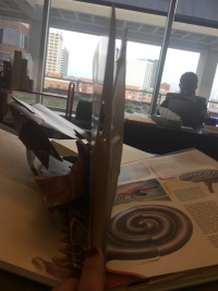
This modern day flap anatomy demonstrates all of the contemporary technologies used to create these books. The left image portrays the complexity of the inside of one page, specifically, two adhered sheets with the middle space used for different flaps and tabs. The image on the right displays the immediate pop up of flaps upon opening to the page. These technologies allowed for cheaper mass-production, as well as the feasibility of sharing flap anatomies, since the limitations of delicacy and easy wear and tear no longer existed, and costs had decreased. Due to the new practicality of these books, flaps started to shift from their original, biological purpose towards one more creative and leisurely: children’s books. [12]
In the modern era, many individuals are familiar with the pop-up castles or 3D dragons in fictional story books. Once printers mastered the technology for flap anatomies, the purpose of the technology was able to expand to various uses, rather than just biological. Additionally, authors saw a market for a creative use of these technologies, and believed they would benefit communities in more than just a medical fashion. This was when the public saw a gradual shift from mostly “flap anatomies” to the emergence of “pop-up books.” Pop-ups in children’s novels now is an entire new genre, in which many individuals have won awards for their work. Robert Sabuda, New York Times Best-Selling Author, is famous for his recreations of past tales, such as Alice in Wonderland, and publishing them using pop-up technologies. [13] These books are still extremely common and can be found in libraries around the world.
For the past six centuries, flap anatomies have revolutionized the way we read, understand and learn. Specifically, they transformed the way in which the public perceives the human body, how doctors teach, and how medical students interpret information. Further, a general audience was able to better understand biological functions, rather than a niche group of medical professionals, which lead to increasing medical innovations and thought. Additionally, emphasis on biological differences between men and women impacted the ways in which women were perceived, as well as helped to define specific gender roles within society. As the technology became more concrete and easily produced, flap anatomies also morphed into pop-up books, which spread to an even larger audience and served as a beloved childhood genre. Overall, using both sight and touch to engage readers maximized understanding, readership and enjoyment of the literature. The impact of this technology is unquestionable, and continues to play an extremely relevant role in the current era.
Notes
- ↑ Gardiner, Bryan. “How Flap Illustrations Helped Reveal the Body's Inner Secrets.” Atlas Obscura, 6 Jan. 2017, www.atlasobscura.com/articles/how-flap-illustrations-helped-reveal-the-bodys-inner-secrets
- ↑ “Anatomy.” Duke University Libraries , exhibits.library.duke.edu/exhibits/show/anatomy/anatomy/intro
- ↑ “Anatomical Fugitive Sheets Printing, Prints and the Spread of Anatomy in the Sixteenth Century.” Https://Europepmc.org/Backend/Ptpmcrender.fcgi?Accid=PMC2531062&Blobtype=Pdf
- ↑ René Descartes, De Homine, 1664, Netherlands Leiden, Rare Book & Manuscript Library, University of Pennsylvania, Photo by Taylor Freeman, October 2nd, 2018
- ↑ “Anatomical Fugitive Sheets Printing, Prints and the Spread of Anatomy in the Sixteenth Century.” Https://Europepmc.org/Backend/Ptpmcrender.fcgi?Accid=PMC2531062&Blobtype=Pdf
- ↑ “Anatomy.” Duke University Libraries , exhibits.library.duke.edu/exhibits/show/anatomy/anatomy/intro
- ↑ “Anatomical Fugitive Sheets Printing, Prints and the Spread of Anatomy in the Sixteenth Century.” Https://Europepmc.org/Backend/Ptpmcrender.fcgi?Accid=PMC2531062&Blobtype=Pdf
- ↑ George Spratt, Obstetric Tables, 1847, Philadelphia: Wagner & M’Guigan, Rare Book & Manuscript Library, University of Pennsylvania, Photo by Taylor Freeman, October 2nd, 2018
- ↑ “Anatomy.” Duke University Libraries , exhibits.library.duke.edu/exhibits/show/anatomy/anatomy/intro
- ↑ “Anatomy.” Duke University Libraries , exhibits.library.duke.edu/exhibits/show/anatomy/anatomy/intro
- ↑ Jonathan Miller, The Human Body, 1983, New York: The Viking Press, Rare Book & Manuscript Library, University of Pennsylvania, Photo by Taylor Freeman, October 2nd, 2018
- ↑ Starr, Michelle. “17th-Century Flap Book Details the Wonders of Anatomy.” CNET, 11 Jan. 2016, www.cnet.com/news/17th-century-flap-book-details-the-wonders-of-anatomy/
- ↑ Robert Sabuda. “Welcome to the Official Website of Robert Sabuda.” Robert Sabuda RSS, www.robertsabuda.com/