File:Brain - CT scan - Metastatic pulmonary adenocarcinoma Case 239 (7603361920).jpg
Jump to navigation
Jump to search
No higher resolution available.
Brain_-_CT_scan_-_Metastatic_pulmonary_adenocarcinoma_Case_239_(7603361920).jpg (512 × 443 pixels, file size: 53 KB, MIME type: image/jpeg)
Summary
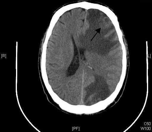
An elderly man appeared in the ER incoherent and cachectic. Brain CT scan was interpreted as showing extensive edema in the left frontal lobe that appears to be related to an underlying 23 mm mass (at arrow). There is a second large area of edema in the left parietal-occipital region. From WikiMedia: https://commons.wikimedia.org/w/index.php?curid=82802824
File history
Click on a date/time to view the file as it appeared at that time.
| Date/Time | Thumbnail | Dimensions | User | Comment | |
|---|---|---|---|---|---|
| current | 07:39, 11 December 2020 | | 512 × 443 (53 KB) | TonyWen (talk | contribs) | An elderly man appeared in the ER incoherent and cachectic. Brain CT scan was interpreted as showing extensive edema in the left frontal lobe that appears to be related to an underlying 23 mm mass (at arrow). There is a second large area of edema in the left parietal-occipital region. From WikiMedia: https://commons.wikimedia.org/w/index.php?curid=82802824 |
You cannot overwrite this file.
File usage
The following page uses this file:
.jpg&oldid=32264)